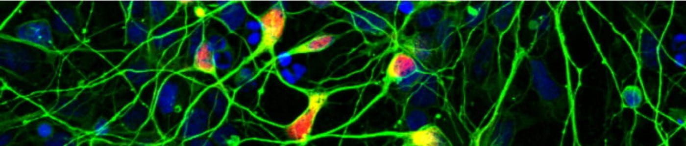
Regional specification of ventral midbrain astrocyte: fine-tuning the inflammatory response.
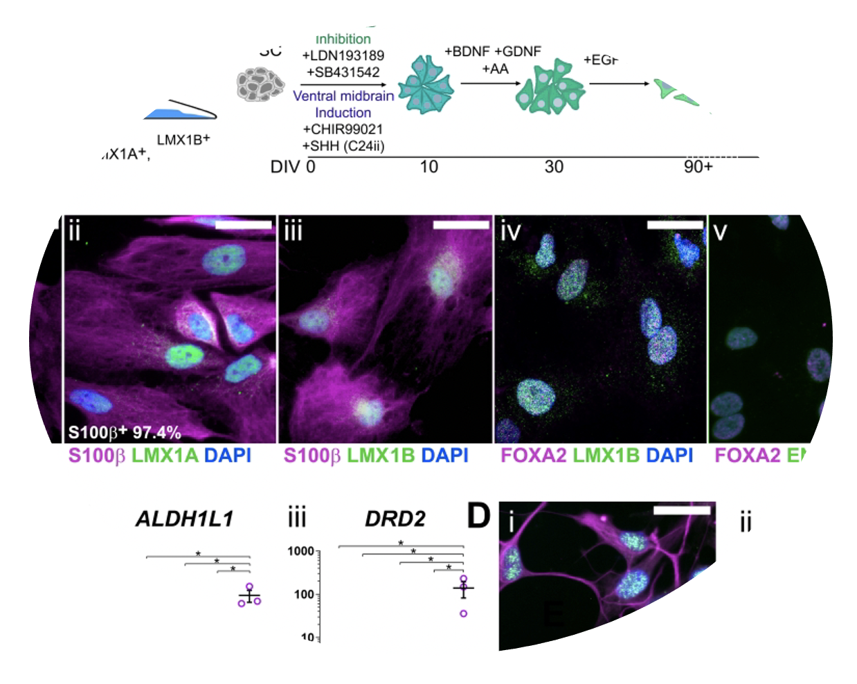 The brain is comprised of multiple distinct regions that perform defined roles through regionally-specified neuronal sub-classes. We are now beginning to appreciate how regional identity influences the functions of other brain cells (glia), principally astrocytes. In order to fully understand how neurons and glia interact with one another during disease-associated stress responses, it is crucial that in vitro models fully consider such regional idiosyncrasies. To address this, we have adapted hiPSC astrocyte differentiation protocols to generate ventral midbrain-patterned sub-types for neuroinflammation research in Parkinson’s. In this study, we use quantitative proteomics to define the inflammatory secretive of these cells, identifying distinct patterns that distinguish these from un-patterned astrocyte populations.
Crompton, LA, McComish, SF, Steward, TGD, Whitcomb, DJ, Lane, JD*, Caldwell, MA* (2023) Human stem cell-derived ventral midbrain astrocytes exhibit a region-specific secretory profile. Brain Comms. https://doi.org/10.1093/braincomms/fcad114
(May 2023)
A new role for ATG8 proteins as transcriptional co-factors.
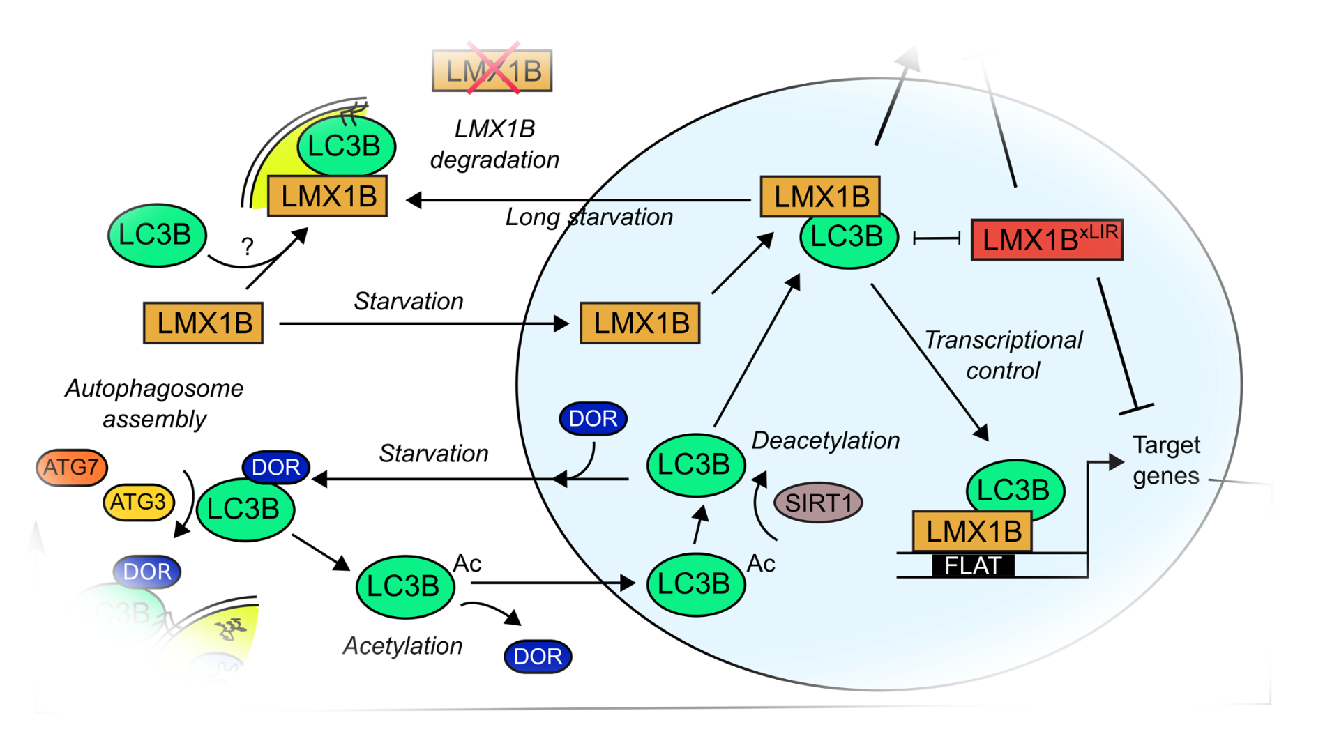
Equisite control of autophagy gene expression during development and stress is needed for proper differentiation and for cell resilience. In the dopaminergic neurons of the human midbrain (“mDANs”) the transcription factors LMX1A and LMX1B are crucial determinants of differentiation and stress resilience, and it is thought that declining expression of either could contribute to mDAN loss in Parkinson’s. Intriguingly, as well as controlling developmental gene expression, these transcription factors also regulate expression of a number of core autophagy (ATG) genes. In this study we identify a new regulatory interface whereby human ATG8 proteins bind to LMX1A/LMX1B via conserved binding regions to stimulate transcription factor activity. This affords enhanced stress protection in human mDANs in vitro. Thus we present a new role for the ATG8 proteins as regulators of autophagy gene expression for neuronal stress resilience.
Jimenez-Moreno, N, Kollareddy, M, Stathakos, P, Moss, JJ, Anton, Z, Shoemark, D, Sessions, R, Witzgall, R, Caldwell, MA, Lane, JD (2023) ATG8-dependent LMX1B-autophagy crosstalk shapes human midbrain dopaminergic neuronal resilience. Journal of Cell Biology 222 (5): e201910133
(see also: Kournoutis Johansen, T (2023) LC3B is a cofactor for LMX1B-mediated transcription of autophagy genes in dopaminergic neurons. Journal of Cell Biology 222 (5): e202303008)
(May 2023)
The ATG5 interactive highlights how autophagy engages with the endocytic system.
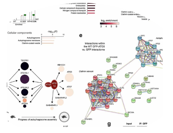 Autophagosomes are transient organelles that are formed de novo in response to stress triggers. Increasingly, we are becoming aware of how regulators of the autophagy pathway engage with other recognised players in distinct membrane trafficking pathways. In order to find novel regulators at the autophagosome assembly stage, we compared interactomes of wild-type and conjugation-deficient GFP-ATG5 using quantitative SILAC-based proteomics. From this, we identified a number of known regulators of the endocytic pathway, and describe how some of these impact of autophagosome assembly and the surface proteome.
Baines, K, Yoshioka, K, Takuwa, Y, Lane, JD (2022) The ATG5 interactome links clathrin-mediated vesicular trafficking with the autophagosome assembly machinery. Autophagy Reports https://www.tandfonline.com/doi/full/10.1080/27694127.2022.2042054
(April 2022)
Autophagy and chondrocyte development: fish find feeding frustrating.
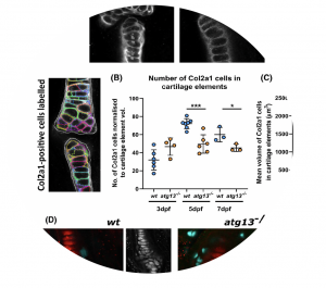 Autophagy plays important roles during the differentiation of various cells and tissue types. Using an atg13 CRISPR knockout zebrafish line established by Martina Worth and Sharon Tooze, we show that absence of autophagy in zebrafish embryos causes early death (17 dpf), with gross phenotypical changes noted around 5 dpf (notably, lack of swim bladder inflation). On close inspection using light and electron microscopy, we observed accelerated chondrocyte maturation and hypertrophy, effecting jaw development. Contributing to the demise of the fish is an inability to open their mouths sufficiently, effecting their feeding post yolk sac exhaustion.
Moss, JJ, Wirth, M, Tooze, SA, Lane, JD, Hammond, CL (2021) Autophagy coordinates chondrocyte development and early joint formation in zebrafish. FASEB J. https://doi.org/10.1096/fj.202101167R
(October 2021)
Developing tools for MitoCRISPR: is type V Cas12a the answer?
 The ability to target mitochondrial DNA (mtDNA) for research and in the clinic is extremely desirable. Attempts using CRISPR technology to target the mitochondrial genome have so far failed or lack corroboration. In this study, we target different CRISPR effectors to mitochondria and apply mitochondrial targeting strategies for g/crRNA import to determine suitability of possible mitoCRISPR systems.
,,, H, , MW,, MD, LVariability in mitochondrial import, mitochondrial health and mtDNA copy number using Type II and Type V CRISPR effectors. Journal of Cell Science
(August 2020)
A SNX4:SNX7 heterodimer controls ATG9A trafficking and autophagy
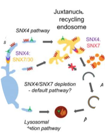 The sorting nexins (SNXs) play crucial roles during the sorting of endocytotic cargo for final destinations within the cell. In yeast and mammalian cells, SNXs are also implicated in the autophagosome assembly process. We show that SNX4 in heterodimer with SNX7 assist in ATG9A mobilisation during autophagy to establish productive assembly sites.
, Z,, VMA,, B,, C,, N, , P,A heterodimeric SNX4:SNX7 SNX-BAR autophagy complex coordinates ATG9A trafficking for efficient autophagosome assembly. Journal of Cell Science
(June 2020)
A new hiPSC dopaminergic neuronal culture system for Parkinson’s autophagy research
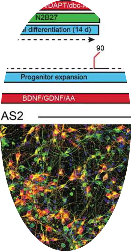 Autophagy cytoplasmic quality control is an important protective mechanism against cellular stress. With evidence linking autophagy dysfunction to the death of neurons in Parkinson’s (the dopaminergic neurons in the midbrain), reliable systems that model Parkinson’s-linked neuronal stress and autophagy are needed. In this paper we document our work towards a reliable culture system using human induced pluripotent stem cells (hiPSC) that produces high numbers of midbrain dopaminergic neurons in a format amenable for autophagy research.
Stathakos, P, Jiménez Moreno, N, Crompton, L, Nistor, P, Badger, J, Barbuti, P, Kerrigan, T, Randall, A, Caldwell, M, Lane, JD (2020) A monolayer hiPSC culture system for autophagy/mitophagy studies in human dopaminergic neurons. Autophagy https://doi.org/10.1080/15548627.2020.1739441
(Feb 2020)
AKT control of apoptosis – a mathematical modelling approach
 The molecular interactions that underpin apoptosis signalling are complex and broad. Using mathematical modelling, we have rationalised these interactions to identify steps and/or protein species that dominate cell death decision making. We identify KT as a highly interconnected mediator of these decisions, and report in vitro and in silicons how AKT regulation coordinates the apoptosis response. Congratulations Matt and Joanna!
Anderson, MW, Moss, JJ, Szalai, R & Lane JD (2019) Mathematical modelling highlights the complex role of AKT in TRAIL-induced apoptosis of HCT116 colorectal carcinoma cells. iScience 12:182-193
(Feb 2019)
Autophagy controls astrocyte-mediated neuronal DNA damage following metal nanoparticle exposure
All the hard work has been worth it! A long-standing collaboration with Maeve Caldwell (Dublin) is now published at Nature Nanotechnology. Lucy, Petros and Natalia are all co-authors in the exciting study that implicate astrocytes and autophagy in neuronal toxicity during nanoparticle exposure across a placental barrier model.
Hawkins SJ, Crompton LA, Sood A, Saunders M, Boyle N, Buckley A, Minogue AM, McComish S, Jiménez-Moreno N, Cordero-Llana O, Stathakos P, Kelly S, Lane JD, Case CP & Caldwell MA (2018) Nanoparticle induced neuronal toxicity across placental barriers is mediated by autophagy and dependent on astrocytes. Nature Nanotechnology doi:10.1038/s41565-018-0085-3.
(2nd April 2018)
siRNA screens for ER-mitochondrial contact sites in autophagy implicate Ca2+ exchange a key mediator
 ER-mitochondrial contact sites are key mediators of cellular calcium signalling. They are thought to play important roles during autophagy by providing platforms for the assembly of the early autophagy machinery. By carrying out parallel screens for factors involved in ER and mitochondrial dynamics and connectivity, we have found that different regulators of Ca2+ exchange contribute to autophagy (SigmaR1) and mitophagy (IP3Rs). This work is published in the International Journal of Molecular Sciences.
Mitochondrial OPA1 cleavage is needed for efficient Parkin-mediated autophagy in OXPHOS-dependent cells

The regulation of mitochondrial dynamics is an essential control for mammalian mitophagy. We showed that in cells dependent on mitochondrial oxidative phosphorlyation for energy, mitochondrial dynamics are altered to prevent mitophagy in a Parkin-mediated mitophagy model system. Probing the molecular control of this key regulatory pathway, we found that OXPHOS-dependent cells resist stress-induced mitochondrial fragmentation by resisting OMA1-dependent OPA1 cleavage at the inner mitochondrial membrane. We also found that DRP1 activity is suppressed under these conditions, contributing to the block in mitochondrial fission. This work is published in the Journal of Cell Science.
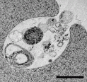
Autophagy and organelle clearance in human erythroid cells
During erythropoiesis, all organelles are cleared from the cytoplasm resulting in reticulocytes and erythrocytes that are devoi d of internal membrane structures. Using human erythroid precursors differentiated from adult stem cells, we reported the timing of these changes in organelle structure and abundance by measuring organelle content using high pressure freezing electron microscopy. Using a lentiviral delivery system to overexpress dominant negative autophagy regulators in erythroid precursors, we showed that the ATG4B plays a role during late stages of erythroid autophagosome maturation. Late stage erythroid cells expressing C74A ATG4B retain swollen amphisomes that contain undegraded material. This suggests that regulated delipidation of LC3 is needed for fusion with the lysosomal compartment. This work is published in Autophagy.
Caspase cleavage of ATG4C and ATG4D reveals latent mitochondrial targeting sequences

The human ATG4 protein family regulate autophagosome assembly by priming then later dilapidating the key autophagy protein ATG8 (LC3). The paralogues ATG4C and ATG4D are cleaved by caspase-3 during stress-induced autophagy, and we previously showed that ATG4D cleavage alters the autophagy response and triggers mitochondrial recruitment and apoptosis (see Betin et al. (2009)). In this follow-up study, we showed that ATG4C and ATG4D each have a mitochondrial import sequence immediately downstream of the DEVD caspase site. Mitochondrial-targeted ATG4D resides in the matrix, disturbs cristae architecture and alters oxidative stress responses to influence cell viability and erythroid differentiation. This work is published in Autophagy.
Sirt3 expression is upregulated in response to oxidative stress

Sirt3 is a mitochochondrial targeted NAD+ dependent deacetylase, implicated in the control of mitochondrial physiology through its capacity to stimulate important metabolic enzymes. We postulated that Sirt3 could play a role in suppressing mitochondrial ROS – a function that would make it an important protective factor in human disease such as Alzheimer’s in which mitochondrial dysfunction contributes. In work published in PLOS one we show that Sirt3 expression correlates with the progression of Alzheimer’s disease in humans and in a mouse model, and that acute stimulation of mitochondrial ROS in vitro stimulates Sirt3 expression. Importantly, we find that overexpression of Sirt3 in primary hippocampal neurons protects against ROS-induced cell death in vitro.
Autophagy contributes to human reticulocyte maturation
We are contributing to the development of protocols for the production of transfusable quantities of human red blood cells from adult stem cells. In this paper we describe a novel approach enabling the generation of mL quantities of packed human reticulocytes. We go on to show that the final stages of reticulocyte maturation involves complex interactions between endocytic an autophagic compartments, establishing nascent exosomes that are subsequently released. These structures label for LC3 and for ER and Golgi membranes suggesting that the final remnants of the erythroid endomembrane system are harboured within. This work is published in Blood.